
In this article we are introducing the MG3 Blue Files for the treatment of Long Mandibular Molar and how the system combines the function of Cutting Efficiency & Flexibility to facilitate the shaping sequence.
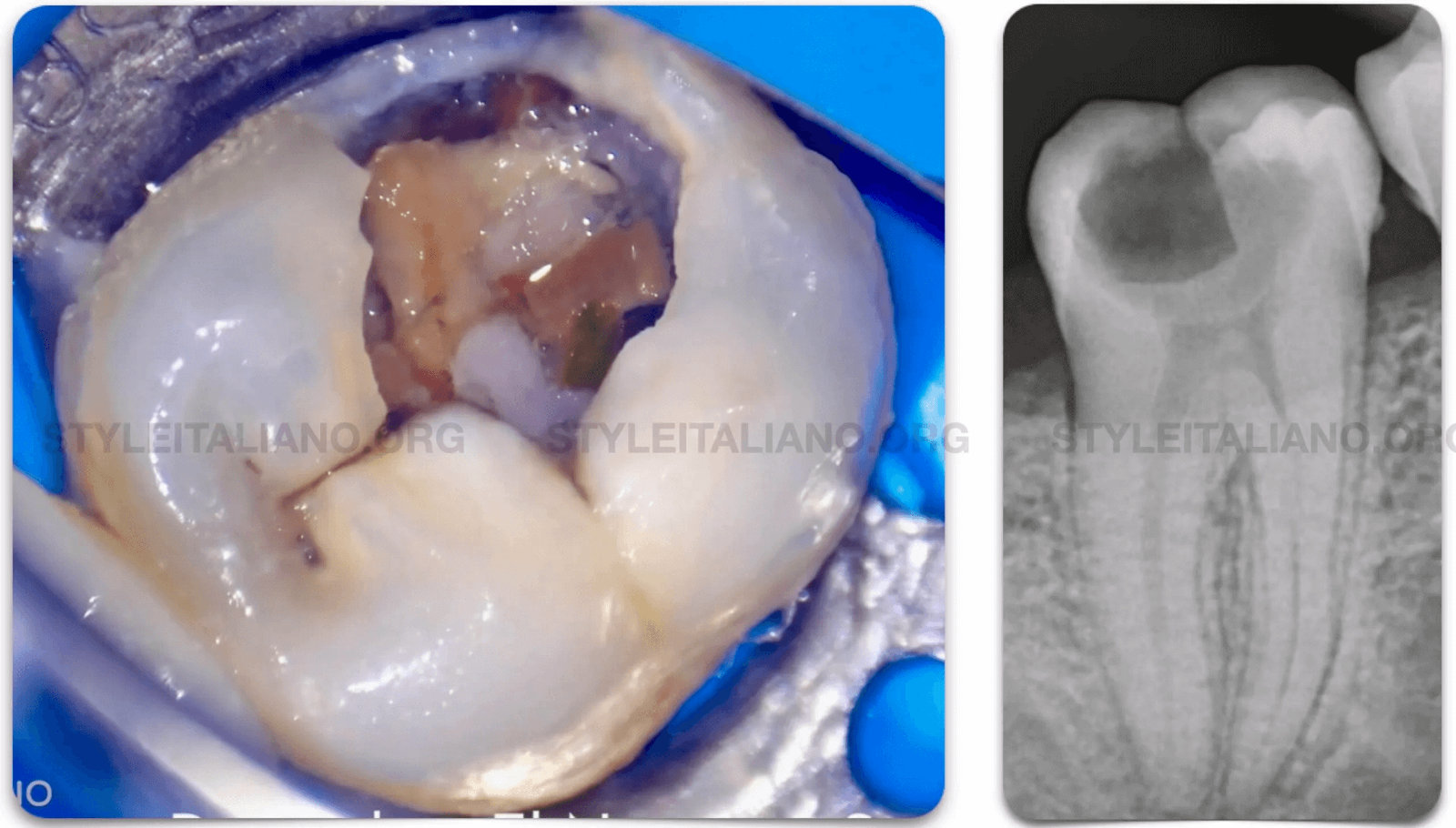

Pre - Op Radiograph , Long Rooted Molar with Coronal Interference in the coronal part of the Mesial System.

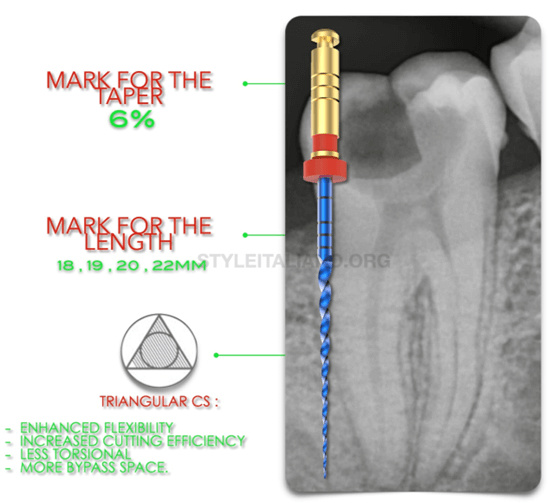
Fig. 2
In the distal Canal we can see that its a large canal , that’s why we can take the advantage of the instrument taper especially the G2 which 25 6% taper to do the flaring and shaping in the same step , also we can shape the canal manulless by using the dual function in the Motor to minimize the number of files and maximize the efficacy.

In the mesial system to unlock the coronal interference we will start mechanical preflaring using the opener which is 20 10% taper and comes in a convex triangle Design which gives the file the needed flexibility beside the cutting efficiency.
The file will be used in the coronal segment with a brushing movement on the outer wall.

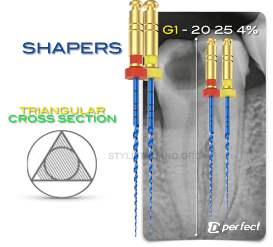
Fig. 4
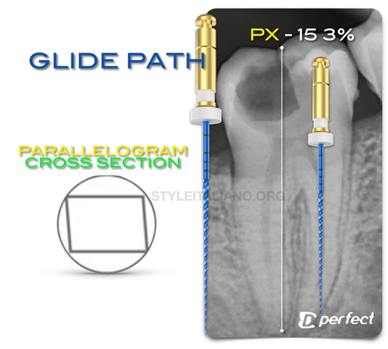
After the mechanical flaring the we are proceeding to the Glide Path Management.
The file of choice will be the navigator which have the features of the martensitic alloy , that makes the file have a high cyclic fatigue resistance and the small mass and taper doesn't allow any unfavorable action of the file like zipping or transportation , lastly the cross section which is PARALLELOGRAM makes the contact between the file and the canal less with move space for Debris evacuation coronally.

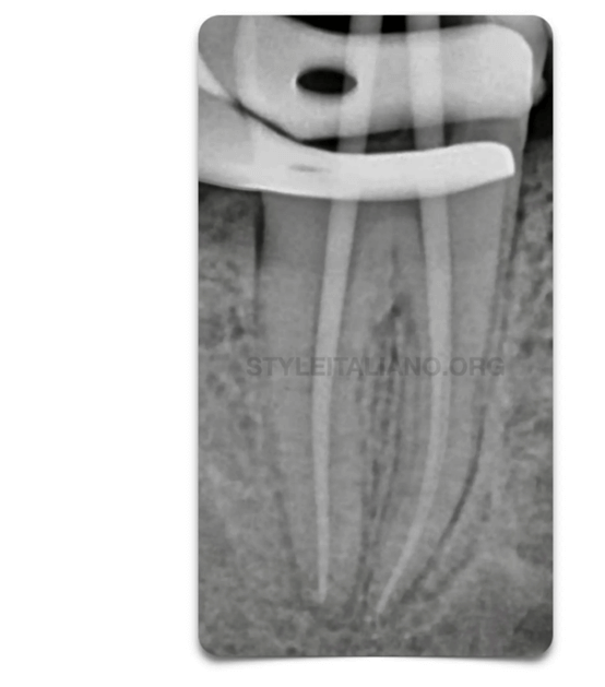
Fig. 5
After the Glide path , the space is now ready for the Shaper G1 ,G2 which is 20 25 4% , the grater benefit of this file is the cross section when it is been combined with the small mass and taper which will aid in shaping of the difficult anatomies , tight canals and also the splits the cross section gives enhanced features for the flexibility and and cutting efficiency.
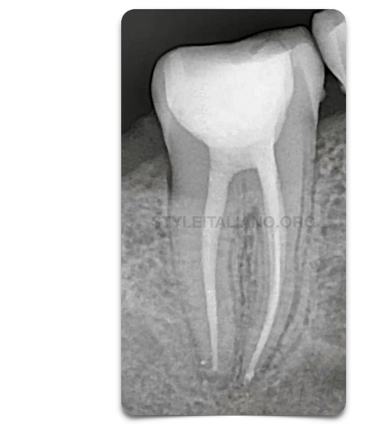
Fig. 6
Master Cone X Ray

The Clinician must have a knowledge about files , kinematics & metallurgy and to choose the best set for every situation , MG3 files and MG3 Blue offers a files for a wide range of Cases and different situation.
1. Endodontic Facts, American Association of Endodontists. Available at: http://www. aae.org/about-aae/news-room/endodontic-facts.aspx. Accessed June 1, 2015.
2. Borges AH, Bandeca MC, Tonetto MR, et al. Portland cement use in dental root perforations: a long term followup. Case Rep Dent 2014;2014:637693.
3. TsesisI,FussZV.Diagnosisandtreatmentofaccidentalrootperforations.Endodontic Topics 2006;13:95–107.
4. Tsesis I, Rosenberg E, Faivishevsky V, et al. Prevalence and associated periodontal status of teeth with root perforation: a retrospective study of 2,002 patients’ medical records. J Endod 2010;36:797–800.
5. Gorni FG, Gagliani MM. The outcome of endodontic retreatment: a 2-yr follow-up. J Endod 2004;30:1–4.
6. Clauder T, Shin S-J. Repair of perforations with MTA: clinical applications and mechanisms of action. Endodontic Topics 2006;15:32–55.
7. Kvinnsland I, Oswald RJ, Halse A, Gronningsaeter AG. A clinical and roentgenological study of 55 cases of root perforation. Int Endod J 1989;22:75–84.


Whether you are a beginner or an expert, this event will give you the opportunity to learn and progress

Product consultant one-on-one service
Free product solutions
Professional team meticulous answer


© 2024 dental-perfect.com. All Rights Reserved.Shenzhen Perfect Medical Instruments Co. Ltd


