


Root Canal Retreatment of Maxillary Second Premolar with Double Curvature and Two Separated Instruments
• The patient was referred to our clinic due to a fractured upper posterior tooth.
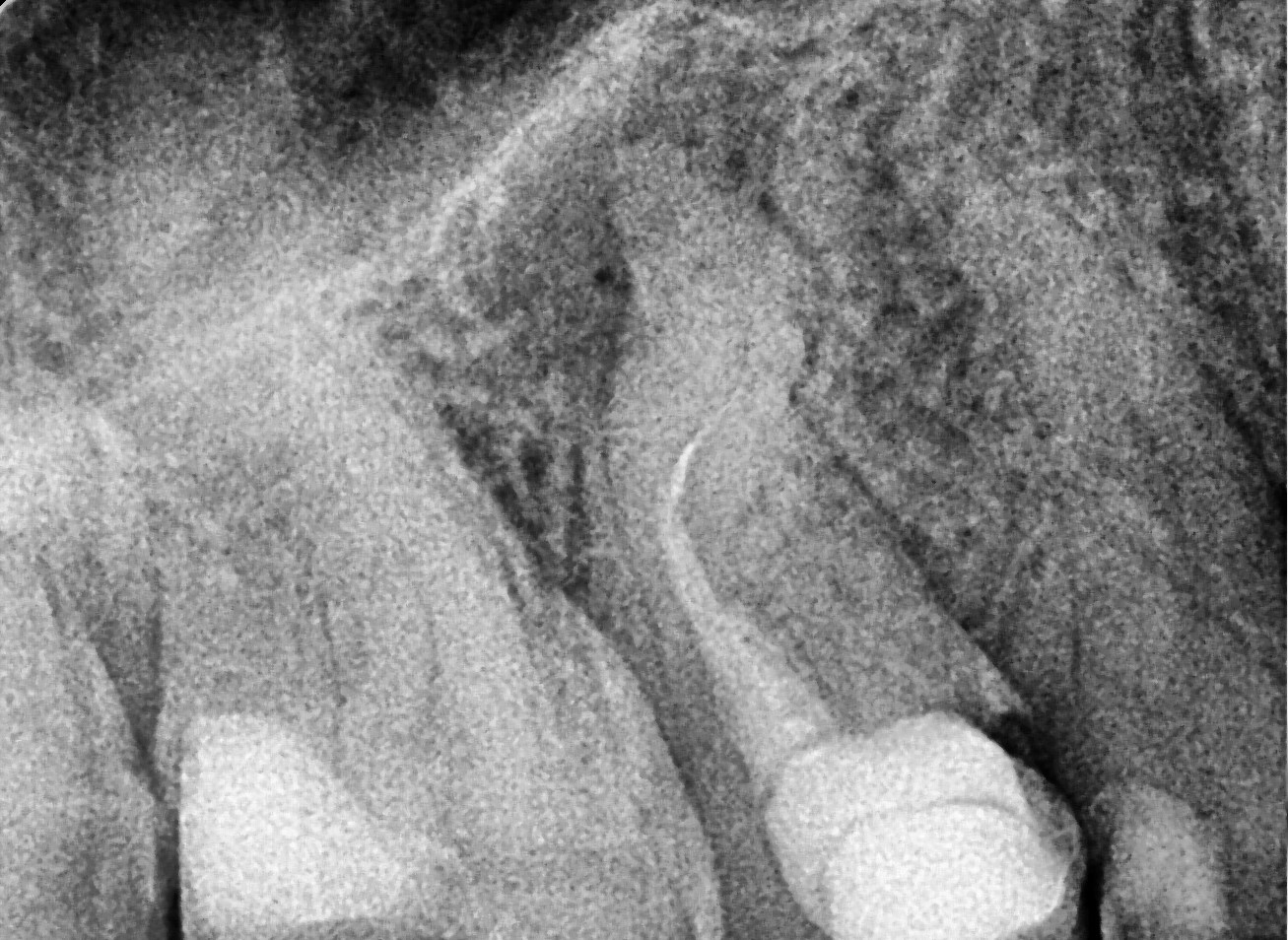
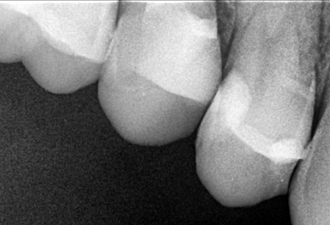
Clinical and radiographic examination revealed a short root filling and the presence of fractured instruments (image 1).
• After rubber dam isolation, treatment was initiated.
The coronal gutta-percha was removed using the SuperSystem “Retreat One” (25/06) file and the Perfect Micro H-shaper (image 2)
• In the palatal canal, the 10/07 “Opener” file from the SuperSystem Advanced Kit was used at 250 rpm and 1.5 Ncm torque, progressing to the middle third.
Following irrigation, 17/05 file was used at the same settings to reach the apex.
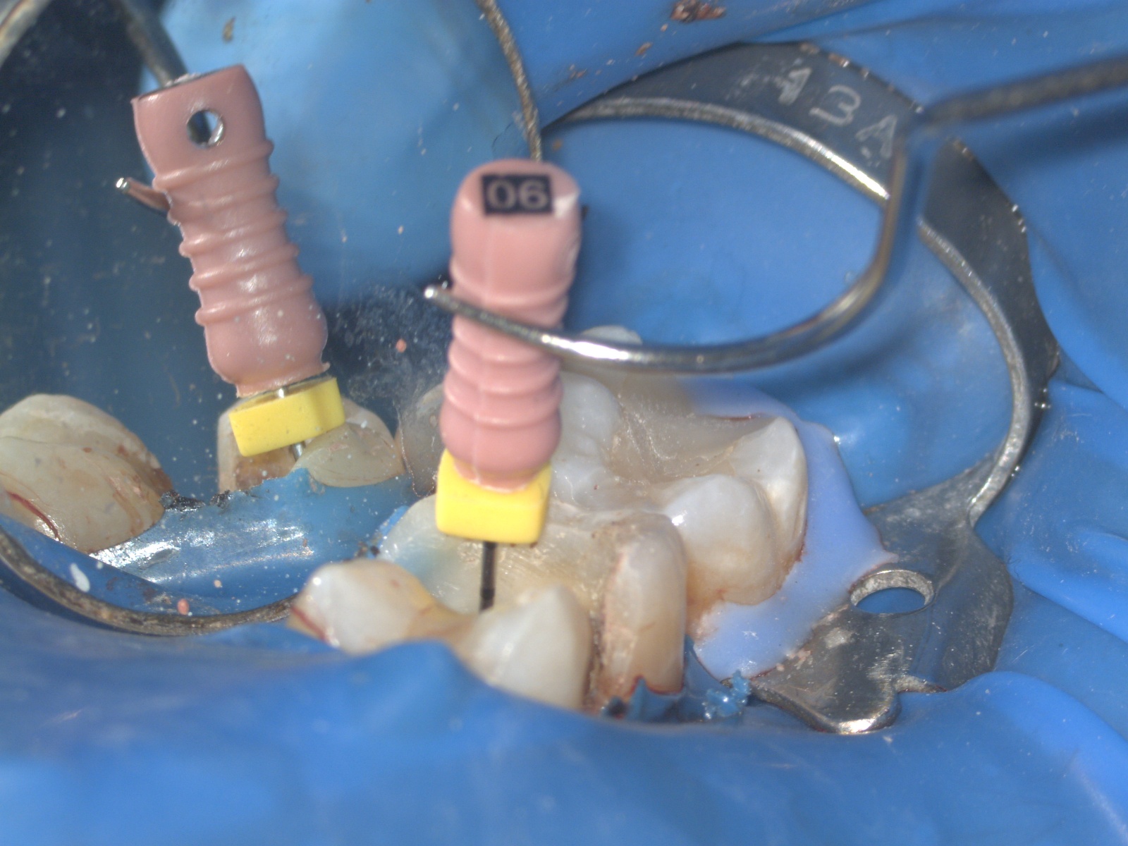
Working length was confirmed using a #06 K-file.
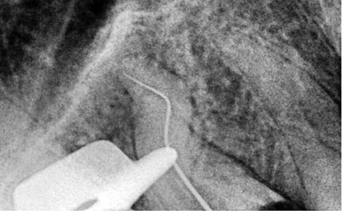
Canal enlargement was completed with the Perfect Advanced Kit 17/05 file (images 3 and 4).
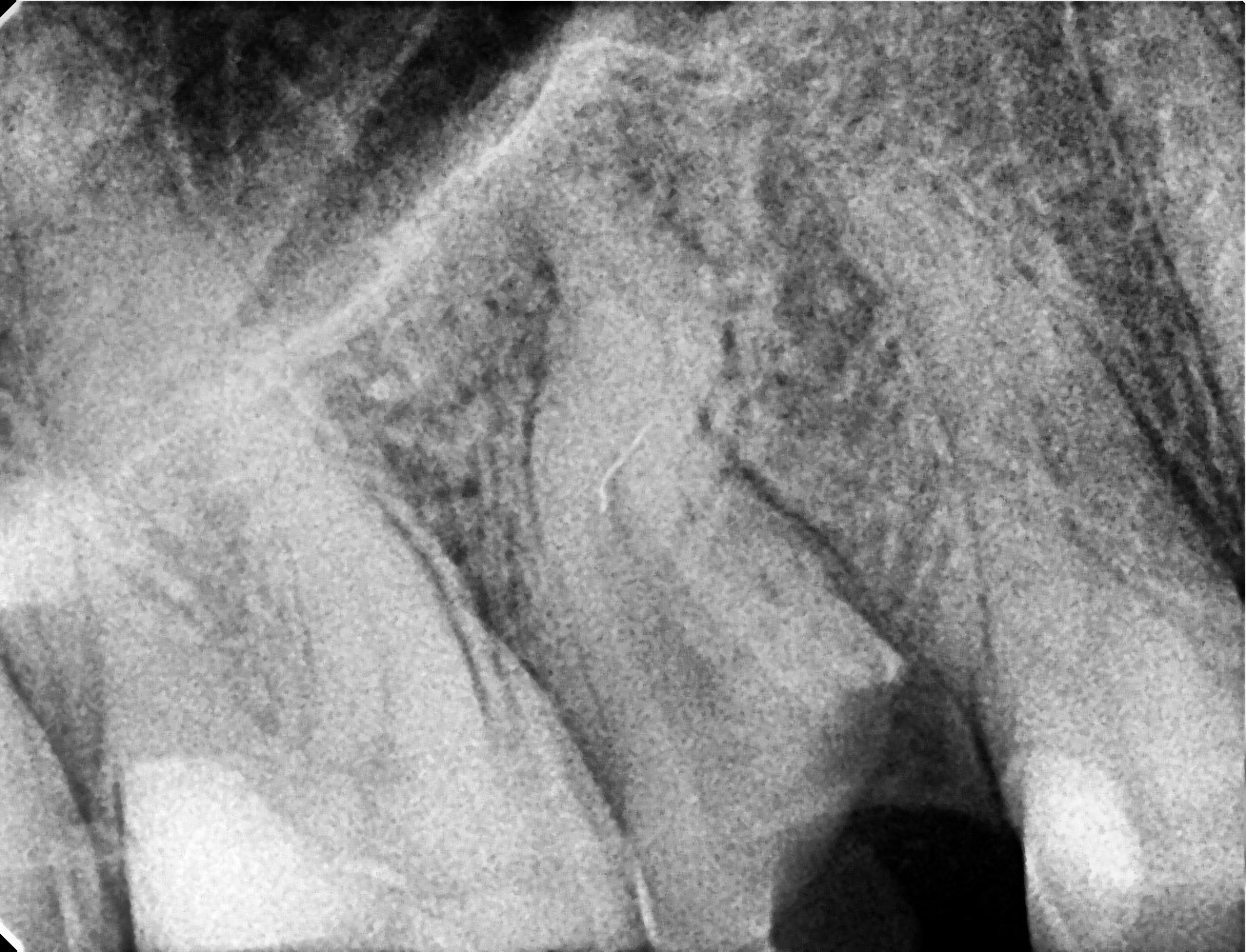
• The buccal canal was dried and debris removed.
Two fractured instruments were detected (image 5), although previously only one was visible due to bucco-palatal superposition on the radiograph.
• The first instrument was removed with ultrasonic tips (images 6 and 7).
The second instrument was loosened with ultrasonic tips and removed using the loop technique (images 8 and 9).
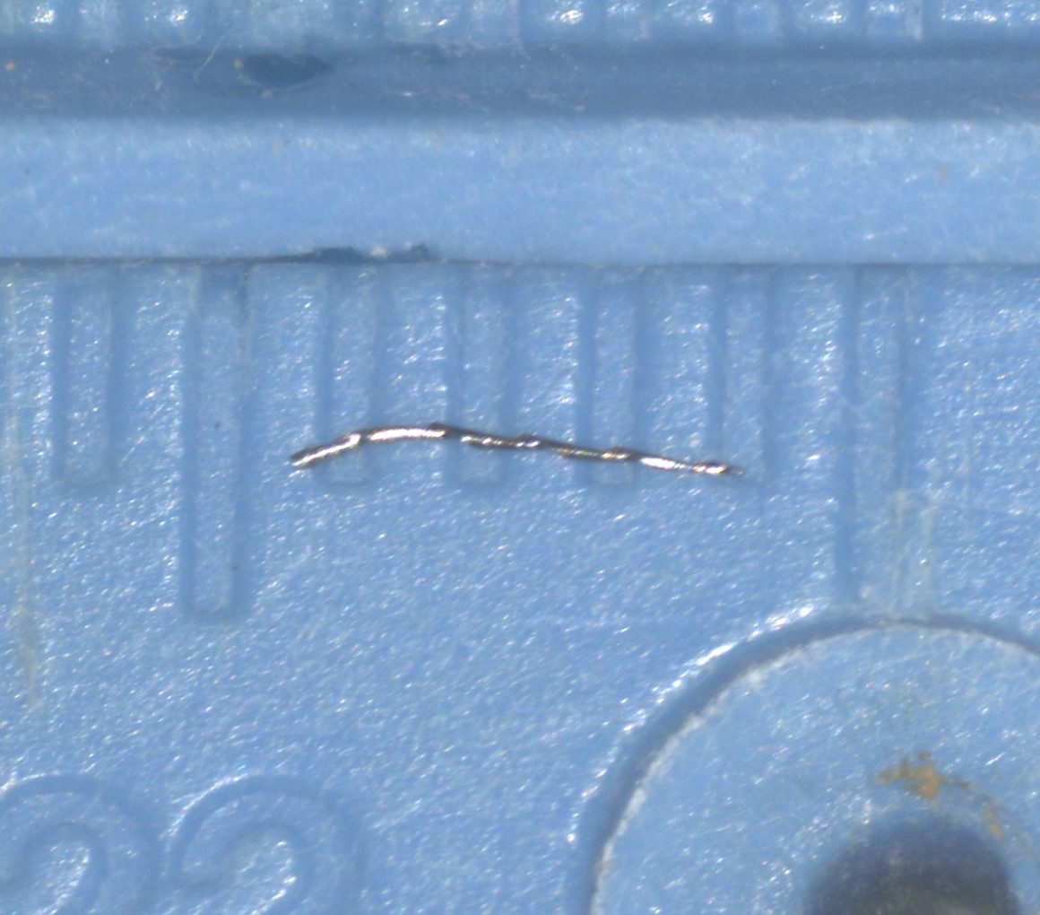
It measured 3.5 mm in length (image 10).
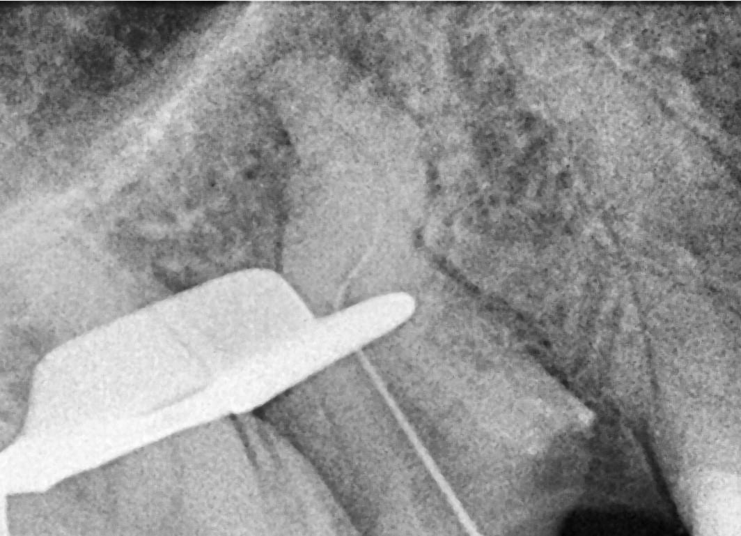
• Attempts to progress with hand files in the buccal canal were unsuccessful in reaching the apex (images 11 and 12).
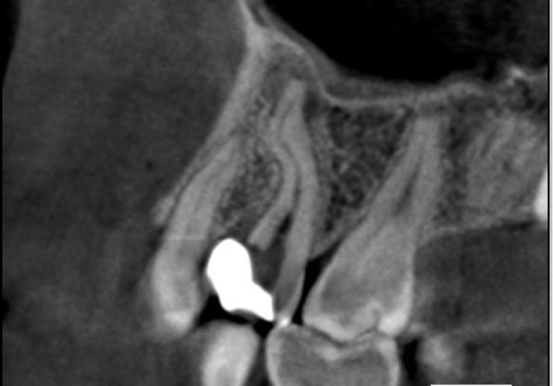
A CBCT scan was taken before the second appointment (image 13).
• According to the scan, the Perfect SuperSystem 10/07 and 17/05 files were pre-curved mesio-distally to match the canal curvature.
Once light resistance was felt, files were operated to perform canal preparation.
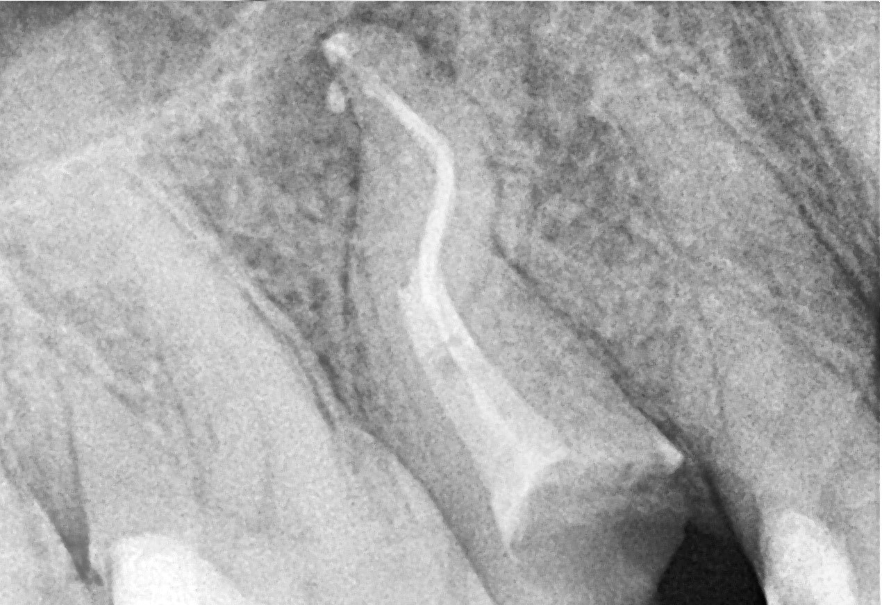
Using the crown-down technique, apical patency was achieved with the 17/05 file (image 14).

• Due to significant canal curvature, long, thin plastic sonic tips were used for irrigation activation (image 15).
• The canals were dried with sterile paper points (image 16).
Obturation was done using bioceramic sealer and gutta-percha (image 17).
• The canal orifices were sealed with high-filler composite (image 18).
The patient was referred to their dentist for indirect restoration (image 19).


Whether you are a beginner or an expert, this event will give you the opportunity to learn and progress

Product consultant one-on-one service
Free product solutions
Professional team meticulous answer


© 2024 dental-perfect.com. All Rights Reserved.Shenzhen Perfect Medical Instruments Co. Ltd


